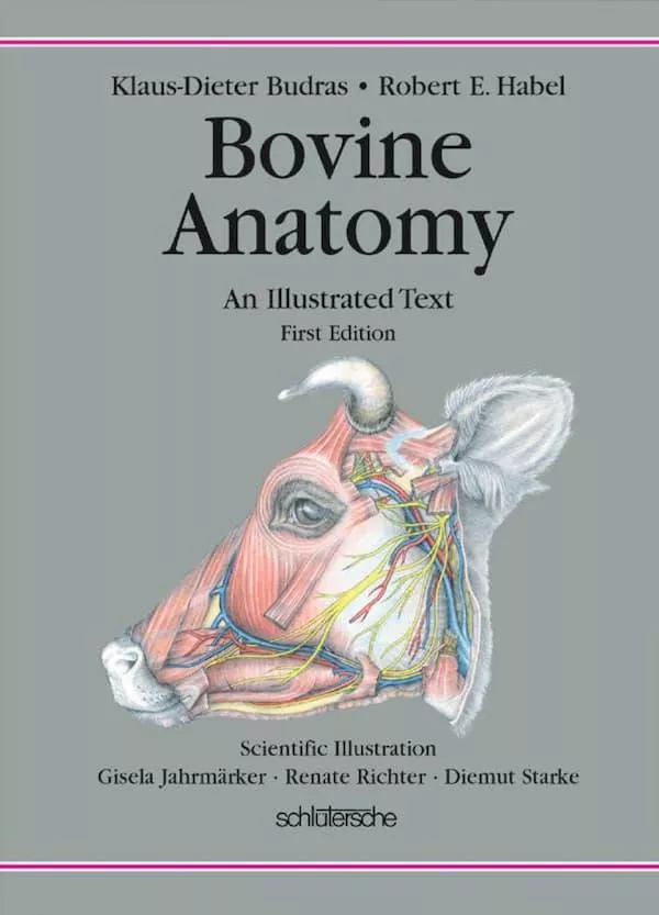


This combination of topographic color atlas and concise textbook of Bovine Anatomy is the third volume of a series on the anatomy of domestic mammals. The first edition of the Atlas and Textbook of the Anatomy of the Dog appeared 20 years ago. It was followed 12 years ago by the second volume, the Anatomy of the Horse. In several German and foreign language editions they aroused world-wide interest. Therefore our next project was an Atlas and Textbook of Bovine Anatomy following the proven model and thereby closing a pre- viously existing gap: no comparable work on bovine anatomy was available. The special features of the ox are presented to students in a well-grounded survey of topographic anatomy. Special anatomy is summarized as brief data in tables of muscles, lymph nodes, and nerves, with references to the corresponding pages in the text. Comparative anatomy is addressed through references to the horse and dog. In addi- tion the text-atlas is intended to provide a valuable introduction to the Anatomy of the Living Animal. The authors were concerned with the preparation of a clear and graphic reference book of important anatomical facts for veterinarians in practice and research as well as anyone interested in morphology. This book can also serve as a dictionary of English anatomical nomenclature illustrated in color. An appendix on Applied Anatomy, included in the first and second volumes of the series, was omitted from this edition. Because of its extraor- dinary relevance for the practical instruction of students it will be provided in the next edition.
Our work on the ox has an unexpected urgency for three reasons: 1. Specialized textbooks for each individual species are required for cur- riculum revision with the trend to premature specialization and the accompanying formation of species-specific clinics. 2. In the present time of economic and social change, new diseases like bovine spongiform encephalopathy (BSE) attain enormous importance through their catastrophic effects. To determine the neuronal pathways of infection, including the autonomic nervous system, and the lymphatic system, and to judge the risk of noxious substances in the nervous system and in many organs of the body cavities, a graphic survey of bovine anatomy is necessary. 3. A licensed veterinarian is legally qualified to serve in a wide variety of positions: in private practice with small mammals, birds, horses, ruminants, and swine; in public health work to prevent transmission of diseases of animals to man; in govern- mental control of diseases of livestock; and in teaching and research with many species of experimental animals. To maintain public con- fidence in the profession, students should be required to master the basic as well as clinical sciences for food animals. This places high demands on teachers and students because a very broad and important body of information must be transmitted even though our teach- ing time has undergone an ill-advised reduction. Nevertheless, we are forced to accept the challenge, even with our compressed text-atlas, to reach the intended goal – to cover a huge amount of subject matter in the short time available.
This English edition is the responsibility of Professor Habel. His translation and scientific engagement in the production of this atlas and the writing and revision of many chapters are his personal service. His collaboration in the community of authors is a great enrichment.
Our thanks are due also to Prof. Dr. Dr. h.c. Simoens (Ghent) for his contributions of text and illustrations on the eye of the ox, to Prof. Dr. Dr. h.c. König (Vienna) for his article on the mammary glands, and to Prof. Dr. Dr. h.c. mult. Liebich (Munich) for his collaboration on the article, “Female genital organs.” Coauthors Dr. Wünsche, Dr. Buda, PD Dr. Bragulla, and Dr. Mülling also had their part in the completion of the book. We had additional professional support from Professors Dr. Berg (St. Kitts, West Indies), Dr. Böhme (Berlin) and Dr. Hashimoto (Sapporo). The many suggestions and the completion of many separate tasks on this atlas by the scientific, student, and technical coworkers of our Berlin Institute (see the list of coworkers) were a great help.
Finally, without the prodigious effort of our excellent artists, Renate Richter, Gisela Jahrmärker, and Diemut Starke, the Atlas in its pres- ent form would be inconceivable. Mrs. Poersch deserves thankful recognition for her careful computer composition, and the coworkers Mrs. Claudia Nöller and Mr. Thilo Voges for the preparation of subjects to be illustrated, together with computer processing, and for mak- ing the Index. Our thanks are also due to the Schlütersche Verlag, Publisher and Printer, Hannover, and especially to Dr. Oslage for always providing support and understanding cooperation in the development of this book.
The provisional completion of our common effort offers the originator and editor, after 30 years of persistent work, the opportunity for a brief reflection. The enormous expense for the production of a book, together with the revision and improvement of many new editions, and the necessity of intensive anatomical preparation of subjects for illustration, were at first greatly underestimated. After overcoming many challenges, the dominant emotion is the joy of an unexpected success that came about through fruitful collaboration with the clos- est coworkers of our Berlin Institute, with the student body, with the readers, and with German and foreign colleagues across national and continental borders. The experience gained thereby is of inestimable value. The editor feels richly rewarded by the achievement of a pro- fessional life-work.